
Occlusal X-Ray
Occlusal X-ray is an intraoral X-ray that utilizes a phosphor plate, which is placed inside the patient's mouth and retained with their teeth.

How is an Occlusal X-ray Performed?
The patient is placed in a sitting position, with a special phosphor plate placed inside their mouth which they bite down on. The operator gives instructions for the correct positioning of the patient's head and with the contribution of an extraoral Χ-ray device the scan, that lasts milliseconds, is been performed.
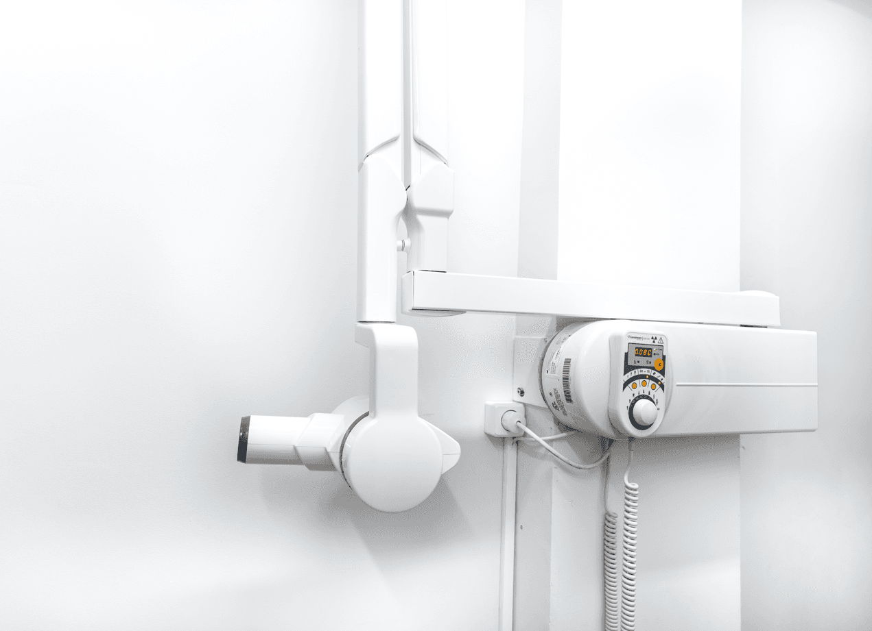
The Imaging System for Occlusal X-ray
The occlusal X-ray is performed by using a dental X-ray equipment, which irradiates a phosphor plate.
Preparation Required
The occlusal examination only lasts milliseconds and doesn't require any special preparation. The only exception is the removal of any removable dental prosthetics the patient may be wearing.
Indications
The occlusal X-ray provides us with valuable information about the upper and lower jaw. Specifically, it is used to detect:
Location of impacted or supernumerary teeth
Nasopalatine cysts
Cysts of the incisive duct
Bone lesions in the palate
Cleft palate
Pathological conditions in the jaw bone
Localization of foreign bodies in the jaws and salivary stones in the ducts of sublingual and submandibular glands

Results Availability
Upon completion of the reception, the printing and receipt of the file are done immediately. The file is then sent electronically, both to the patient and to the attending dentist if requested.

Advantages and Peculiarities
The occlusal X-ray is a unique imaging tool for visualizing anatomical structures of both the upper and lower jaw including the hard and soft palate, incisive foramen, median palatal suture, alveolar bone, nasolacrimal duct orifice, floor of the lower jaw, and beard spines.
While it depicts the anterior teeth of the jaws, it's not the test of choice for imaging posterior teeth.

What Information Does it Provide the Dentist?
The maxillary occlusal image provides information about the hard palate region, the border of the soft palate, the incisive foramen, the median palatal suture, the alveolar bone, and the opening of the nasolacrimal duct.
The mandibular occlusal image depicts the floor of the mouth, the body bone of the lower jaw, and the genial tubercles. Although it can visualize anterior teeth, it's not the preferred method for posterior teeth.
This specialized dental radiography requires knowledge of the anatomical structures of both the upper and lower jaw for accurate execution.


