
Full Mouth - complete series of intraoral X-rays
The complete series of intraoral X-rays, known as Full Mouth, is a dental radiological examination that is becoming increasingly popular, as it can visualize each tooth separately, without the co-projection of anatomical structures, as is the case with a panoramic X-ray.
Understanding Full Mouth X-ray Series
The Full Mouth X-ray series consists of 14 to 20 intraoral dental X-rays that capture all the teeth in the dental barrier. Each phosphorous fluorographic film displays one to two adjacent teeth, showing the crown, the root, and approximately two millimeters of the alveolar bone.
This examination is especially valuable for new patients, as it provides the dentist with a comprehensive overview of the patient's dental history, current issues, and necessary treatment plan. The Full Mouth X-ray series is a fundamental imaging tool in detecting periodontal disease.
Procedure: How is a Full Mouth X-ray Series Performed?
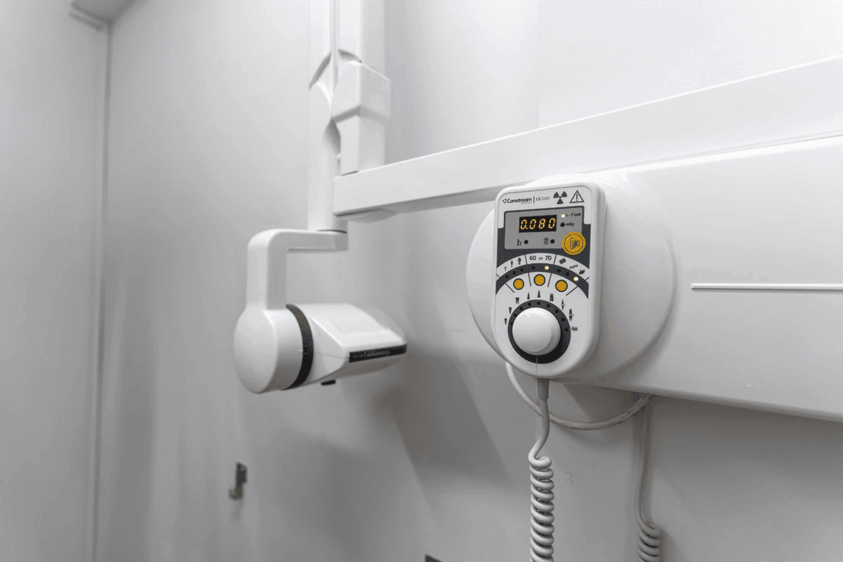
The Full Mouth X-ray series requires no preparation on the patient's part. The patient is seated wearing a protective lead apron, limiting radiation exposure. The operator instructs the patient to bite on the suitable for each type of tooth special retainer holding a phosphorous film the parallel cone technique is applied and the film is exposed to radiation. This procedure is repeated 14 to 20 times to display all the teeth of both barriers.The parallel cone technique eliminates image magnification and projections.
This process is repeated to visualize all teeth, with the use of a special scanner the image is transfered to a computrer. Each X-ray exposure lasts only milliseconds, and the entire examination typically takes around 15 minutes.

Applications of Full Mouth X-ray Series
The Full Mouth X-ray series plays a crucial role in early detection of dental diseases, thus preventing complications. Specifically, it aids in the identification of:
Cavities and dental substance loss
Periapical or apical lesions
Internal or external root resorption
Crown or root fractures
Alveolar bone loss
Periodontal bone loss/disease
Placement of crowns and fillings
Osseointegration of implants
Root canal anatomy and morphology
Pulp anatomy
Evaluation of prosthetic and endodontic root canal therapy

Results and Radiation Exposure
The examination results are immediately available for printing on film or recording on CD. Results can also be sent electronically to the patient and their dentist upon request.
As with all types of x-ray examinations, the Full mouth x-ray series must be performed after a medical referral. To put this in context, everyday presence on Earth's surface exposes us to around 10 micro-Sieverts of radiation daily. Full Mouth X-rays expose patients to roughly 35 micro-Sieverts of ionizing radiation, less than the radiation received on a 7-hour flight.
The radiation dose from a Full Mouth examination is similar to that from a panoramic X-ray, but the lead apron worn during the procedure protects the patient from scattered radiation and dramatically reduses the effective dose received by the patient.
Advantages and Unique Features:
The Full Mouth X-ray series plays a crucial role in early detection of dental diseases, thus preventing complications. Specifically, it aids in the identification of:
Cavities and dental substance loss
Periapical or apical lesions
Internal or external root resorption
Crown or root fractures
Alveolar bone loss
Periodontal bone loss/disease
Placement of crowns and fillings
Osseointegration of implants
Root canal anatomy and morphology
Pulp anatomy
Evaluation of prosthetic and endodontic root canal therapy

Why Choose Dentalray?
At Dentalray, we bring years of expertise and a dedicated team to ensure proper execution of complex diagnostic X-rays such as the Full Mouth series. The expertise required to position X-ray films inside the patient's mouth and utilize dental X-ray retainers to avoid magnification and anatomical structure projection is something we have in abundance.
Our state-of-the-art digital dental equipment and high-resolution dental scanners, coupled with our proficiency, ensure X-rays of high diagnostic value free from artifacts, magnifications, and false images.
At Dentalray, our wealth of experience and exposure to diverse cases equip us to handle even the most challenging situations promptly and effectively. Trust Dentalray for a thorough, precise, and safe dental diagnostic experience.


