
Panoramic X-ray
A Panoramic X-ray presents a comprehensive image, displaying the patient’s maxillary and mandibular oral facial structures across a flat surface. This is an essential diagnostic tool in dentistry, providing valuable insight for treatment planning, particularly in the realm of dental surgery. Panoramic X-rays, however, should only be conducted in specialized diagnostic centers that possess the necessary expertise and sophisticated equipment to produce high-precision results.

Understanding Panoramic Radiography
Panoramic radiography is a radiological examination and a pivotal tool in the dentist's daily clinical practice. Using a minimal dose of radiation, panoramic radiography enables two-dimensional (2-D) imaging of the entire oral cavity. This includes the upper and lower jaws, all teeth, temporomandibular joints, the nasal area, and the maxillary sinuses.
Though the alveolar bone is a hollow structure resembling a horseshoe, panoramic radiography captures it in a flat image, delivering valuable information to the dentist. The scan lasts just a few seconds and is perfectly safe for patients.

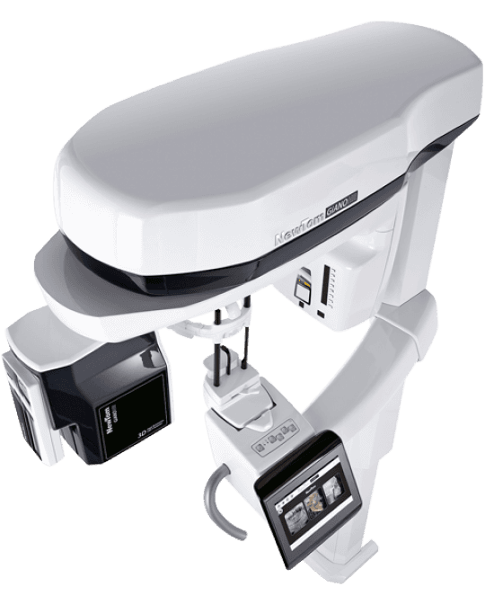
Procedure: How is a Panoramic X-ray Conducted?
The panoramic radiography process does not necessitate special preparation. However, it is important that the technicians are informed if the patient is pregnant. Eyeglasses, jewelry, or other metal objects that may interfere with X-rays must be removed before the examination.
Panoramic radiography is performed with a specialized X-ray device known as an orthopantomographer. This term combines the word 'orthos'—referring the right angle formed by the X-ray beam with the jaw, 'panorama'—indicating the unobstructed view of an object in any direction, and 'tomography'—which pertains to the selective reception of an object free from adjacent and interfering anatomical structures.
The patient is standing upright, biting a special bite block and is immobilized by a head fixation to ensure stillness during the procedure (which lasts a few seconds). The position of the head and body are crucial, as they determine how sharp, clean, and clinically useful the image will be. The X-ray device makes a rotational motion around the patient's head, irradiating the area of the upper and lower jaw. The process is safe, painless, and brief, with the whole procedure taking only a few seconds. Following this, the examination can be printed on X-ray film.
Applications of Panoramic X-ray
Orthopantomography offers a detailed display of all hard tissues in the lower half of the face. It equips the experienced dentist with a comprehensive image of the patient's mouth, significantly aiding in recording and diagnosing dental problems, and in creating a complete, realistic, and feasible treatment plan.
Radiological examination is integral for the dentist, as many diseases of the teeth and jaw bones can only be detected radiographically.
Moreover, it serves as a preventive measure against diseases of the oral cavity, as the dentist can detect potential pathologies in time.
This proactive approach allows the patient to address dental problems at an early stage, minimizing pain and receiving the necessary treatment in a timely manner.
In specific terms, panoramic radiography can reveal various conditions, including:
Anatomical abnormalities or lesions in the upper and lower jaw
Genetic or congenital absence of teeth
Impacted or supernumerary teeth
Location of impacted wisdom teeth and their contact with adjacent anatomical structures
Dental age of children
Cavities in areas not visible during a clinical examination
Existing dental fillings
Bone loss in case of periodontal disease
Periapical inflammation and granulomas
Location of dental implants and assessment of the bone surrounding them
Quality, volume, and regeneration of bone graft
Evaluation of fillings and prosthetic work placement
Radiological examination with panoramic radiography is essential in cases such as:
Injuries to the maxilla and mandible
Medical history of trauma to the head or facial area
Presence of a foreign body in the maxilla-mandible
Tooth fractures
Suspicion of pathological findings in the maxilla-mandible
Impacted teeth or congenital tooth deficiencies
Dysfunction of the temporomandibular joint
Restoration of edentulousness with the placement of osseointegrated implants
Βefore or during orthodontic treatment
Sometimes, a panoramic X-ray alone is not sufficient for a complete diagnosis. In these instances, panoramic radiography is combined with other diagnostic procedures, such as periapical X-rays, cone beam computed tomography of the jaws, tissue biopsy, and puncture.

Results and Radiation Exposure
The printing of the examination on film and its recording on a CD is done immediately. The results can be dispatched electronically to both the patient and the attending dentist upon request.
Radiation surrounds us in various forms. The dose a patient receives during a panoramic X-ray is negligible—it corresponds to the environmental dose one would receive after 12 hours of exposure to a cosmic radiation. Nevertheless, it should always be performed only if deemed necessary by the attending dentist.

Advantages and Special Features
ADVANTAGES
Quick visualization process that takes only seconds
Lower radiation dose compared to other imaging modalities
Comprehensive view of both jaws in a single image
As an extraoral X-ray, it is a simple procedure for the patient but requires expert handling by the operator
Suitable for children aged four years and above, as well as for individuals with disabilities or mobility difficulties
NOTE: Women should always inform the medical team about the possibility of pregnancy as well as breastfeeding.

Why Choose Dentalray?
At Dentalray, we pride ourselves on our extensive experience and specialization in performing panoramic X-rays. We are committed to delivering the best possible service and care to our patients.
Our facilities are equipped with state-of-the-art devices, and our staff is proficient in using cutting-edge software. During any examination or x-ray, we reciprocate our patients' trust by offering high-quality services provided by the best doctors and diagnosticians in the dental radiodiagnostic field.
Dentalray is a highly specialized diagnostic clinic with the exclusive subject of Maxillofacial Radiology
Thus we believe in imparting the right knowledge, ensuring proper placement, and offering guidance to our patients for the best diagnostic results. Panoramic X-rays may be supplemented with complementary X-rays (ex, periapical Χrays) free of charge, depending on each patient's medical condition, to clarify and focus on structures that require further study.
Our staff is always ready to assist you and schedule an appointment according to your needs. During your visit, we consider it our responsibility to inform you about the entire procedure and ensure your comfort. Trust Dentalray for superior dental diagnosis.


