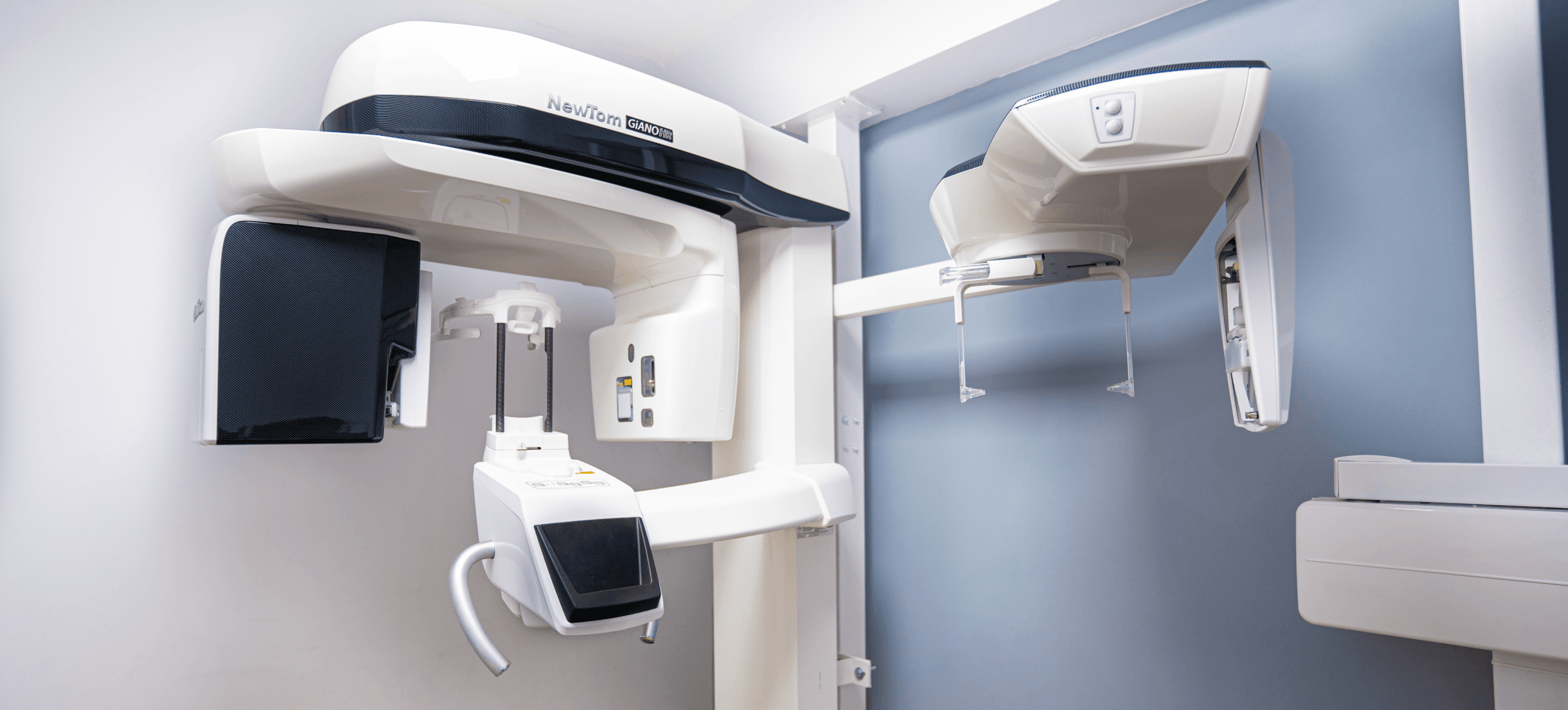
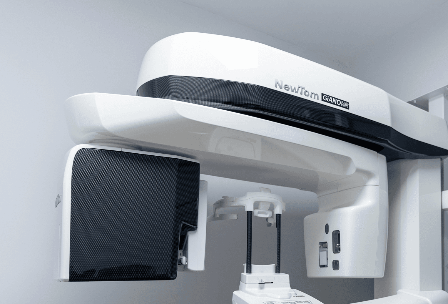
NewTomGiANO HR 2D CEPH
Orthopantomograph
NewTomGiANO HR 2D CEPH
Orthopantomograph
NewTomGiANO HR 2D CEPH
Orthopantomograph
What is NewTomGiANO HR 2D CEPH?
What is NewTomGiANO HR 2D CEPH?
What is NewTomGiANO HR 2D CEPH?
The GianoHR orthopantomograph is an innovative system for obtaining clear and homogeneous panoramic and cephalometric images in an extremely small and flexible device, which offers detailed images for adults and children, further reducing their radiation exposure. The high-sensitivity CMOS sensor and innovative X-ray tube of the GianoHR orthopantomograph ensure ground-breaking image quality and lead to the accurate evaluation of teeth for orthodontic treatment, unerupted dental implants (including fractures – bone abnormalities) and dental prostheses, on both panoramic and cephalometric radiographs.
The GianoHR orthopantomograph is an innovative system for obtaining clear and homogeneous panoramic and cephalometric images in an extremely small and flexible device, which offers detailed images for adults and children, further reducing their radiation exposure. The high-sensitivity CMOS sensor and innovative X-ray tube of the GianoHR orthopantomograph ensure ground-breaking image quality and lead to the accurate evaluation of teeth for orthodontic treatment, unerupted dental implants (including fractures – bone abnormalities) and dental prostheses, on both panoramic and cephalometric radiographs.
The GianoHR orthopantomograph is an innovative system for obtaining clear and homogeneous panoramic and cephalometric images in an extremely small and flexible device, which offers detailed images for adults and children, further reducing their radiation exposure. The high-sensitivity CMOS sensor and innovative X-ray tube of the GianoHR orthopantomograph ensure ground-breaking image quality and lead to the accurate evaluation of teeth for orthodontic treatment, unerupted dental implants (including fractures – bone abnormalities) and dental prostheses, on both panoramic and cephalometric radiographs.

Where is it used?
Where is it used?
Where is it used?
Radiological examination with the GianoHR orthopantograph is considered necessary in cases of patients who:
Start or check their orthodontics treatment
The possibility of having impacted teeth or congenital tooth deficiencies is investigated
They are going to restore edentulousness with the placement of osteointegrated implants or other prosthetics treatments
There is a suspicion of pathological process in the jawbones
Have injuries or a history of an accident in the orofacial region]
Thy have signs of dysfunction of the temporomandibular joint
The possibility of having dental problems such as cavities, lesions, tooth fractures etc


Technical Specifications
Technical Specifications
Technical Specifications
ADVANCED KINEMATIC FEATURES
The specially synchronized kinematics consisting of one rotational motion combined with two simultaneous movements ensures constant magnification in all projections, excellent rectangularity and excellent quality diagnostic images.
FULL CEPHALOMETRIC IMAGING
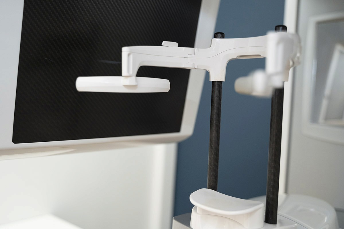
The high-power X-ray lamp and updated positioning system are designed to provide detailed teleradiographic images. The highly sensitive sensor ensures ultra-fast scans to improve patient safety and comfort. The second regulator on the rotating arc facilitates patient access. The use of protectors specially designed for paediatric applications allows the cover of the skull be included in the scan while reducing thyroid exposure.
MULTIPLE PANORAMIC IMAGES (ApT)
Multipan mode creates a total of 5 radiographic images from a single scan. Therefore, the best panoramic of them can be selected for the diagnostic needs of the examination. This function is necessary for the study of complex morphologies. Self-adaptive panorama with ApT (Autoadaptive Picture Treatments) technology provides optimal focusing of the front roots, adaptable to each patient and improves image quality in anatomical area.


